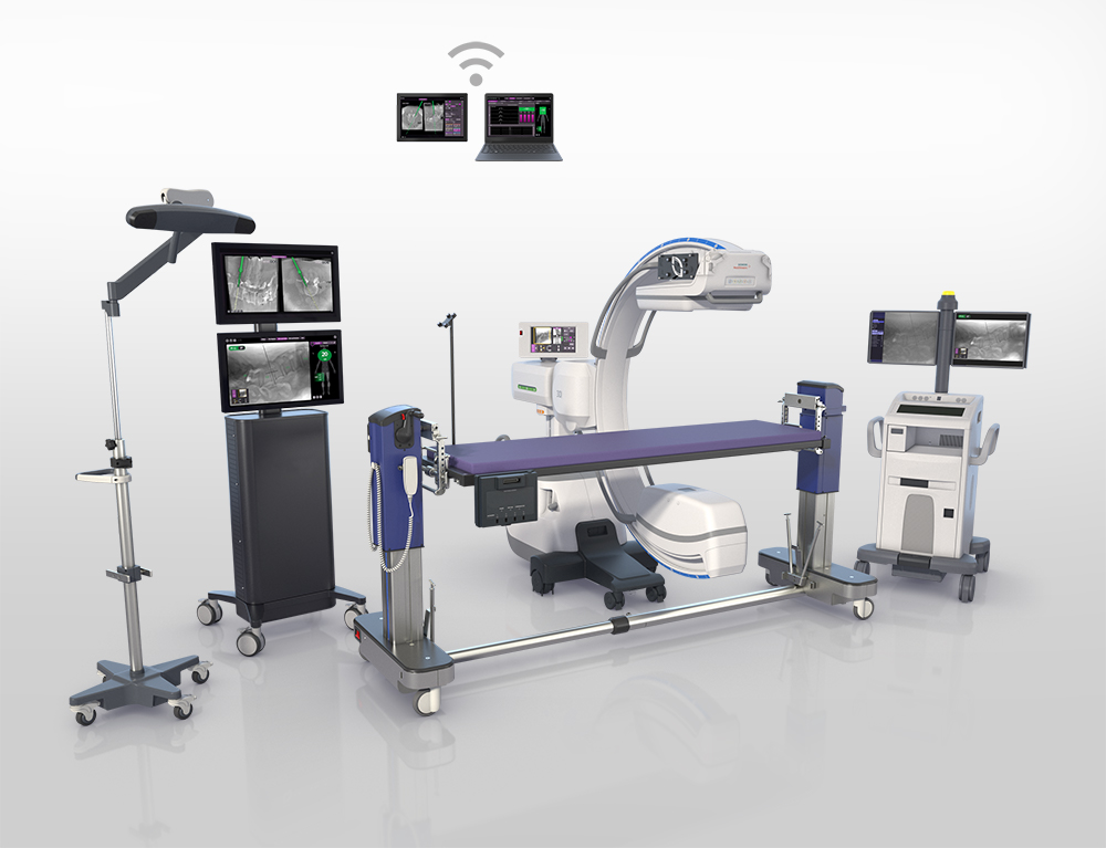
Example of image-guided surgery equipment layout in an operating room. Note the surgical table (purple top) and the fluoroscopy imaging device (C-shaped device adjacent to the operating room table). The fluoroscopy imaging device is used to take x-rays of the patient’s spine. Cameras (located on the horizontal bar on top of the pole with wheels) detect the precise positions of instruments in the operative field and relay this information to computers that then calculate the relative positions of the instruments to the patient’s spinal anatomy based on the pictures from the fluoroscopy device. Monitors then project images for the surgeon to see of the patient’s anatomy that also show the relative position of the surgeon’s instruments. This allows the surgeon to “see” the inside anatomy of the patient’s spine in order to direct instrumentation from the surface.
What is Computer-navigated Spine Surgery?
Your spine isn’t just a bunch of vertebrae stacked upon each other. It’s your body’s central support structure, complete with delicate blood vessels, nerves, and various moving parts. Avoiding trauma to these structures during spinal surgery is our utmost concern. That, and the increased need for precision has paved the way for computer assistance during some complex spine procedures.
Computer navigation can play a passive role by providing real-time information to help the surgeon make critical decisions on the go, or by limiting their movements beyond a certain point. In the fture, this technology may also take on a more active role and actually help perform the surgery!
What is Image-guided Spine Surgery (IGSS)?
Imaging techniques, such as CT scans and fluoroscopy, can be integrated into the system and facilitate pre-operative planning of the surgery. By taking a series of images beforehand, we can display them on the screen and have the surgeon show you the plan for surgery to keep you informed, and with a better peace of mind. Not only that, but these images can also serve as real-time guidance for the surgeon to view and navigate instruments/implants as they’re placed during the surgery.
We can pair these spine images with computer navigation systems. The technology works by using a tracking optical camera to view and select certain body parts as landmarks with infrared markers. These are then transferred to a software that compares them to a model created by the stored CT scan images of the patient’s spine taken during the pre-plan. This can help ensure that the procedure is being carried out in accordance with your individual anatomy.
When is Image-guided Spine Surgery used?
IGSS is being used in many spinal procedures such as the insertion of implants and the decompression of pinched spinal nerves.
For example, in the past thoracic spinal surgery has been faced with many challenges due to the narrow spinal canal and the variable small sizes of the bony pedicles used for stabilizing your thoracic spine. As a result, traditional procedures were met with higher screw misplacments. Use of complex image guidance can help facilitate screw placement for these and other more challenging cases.